The knee is one of the greatest and most complex joints in the body. The knee joins the thigh bone (femur) to the shin bone (tibia). Knee joints comprise two distinct bones. A group of bones that run close to the tibia (fibula) and the kneecap (patella) make up the knee joint.
Annoying knee issues are related to the leg muscles that move the knee joint. Ligaments join the knee bones and give the knee its strength:
- The front cruciate ligament holds the femur back from sliding backward on the tibia (or the tibia sliding forward on the femur).
- The back cruciate ligament holds the femur back from sliding forward on the tibia (or the tibia from sliding backward on the femur).
- Normal and equal assurance ligaments hold the femur back from sliding side to side.
- Two cross-molded pieces of a tendon called the normal and equal menisci to act as protection between the femur and tibia.
- Different bursae, or fluid-filled sacs, help the knee function according to plan.
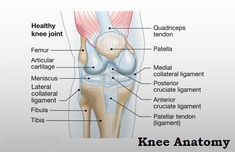
Table of Contents
All About Knee Anatomy
The knee is the most stunning joint in humans.
The knee is a turning joint that is fundamental for weight-bearing and improvement. It includes bones, meniscus, ligaments, and tendons.
The motivation behind the knee is to fulfill different limits:
- Support the body in a standing position without requiring muscles to work
- Helps to reduce weight and heighten the body
- Gives sufficiency
- It comes to fruition as a shield
- Licenses bending of the leg
- Makes walking more useful
- Pushes the body forward
The following sections outline the fundamental elements of knee anatomy.
Bones
The femur (thigh bone), tibia (shin bone), and patella (kneecap) make up the bones of the knee. The knee joint keeps these bones in proper alignment.
The patella is a small, triangular-shaped bone that sits at the front of the knee, inside the quadriceps muscle. Since it gets past significant amounts of tension, it fixes on the thickest layer of a tendon in the body.
Cartilages
There are two sorts of cartilage in the knee:
Meniscus: these circles act as a cushion, or “shield”. So the bones of the knee can move freely without getting in the way of each other. The menisci also contain nerves that help with the creation of balance and assurance of the right weight distribution between the femur and tibia.
The knee has two menisci:
- average – on the internal side of the knee, this is the greatest of the two
- horizontal – outwardly of the knee
Articular ligament: found on the femur, the most elevated place of the tibia, and the back of the patella; it is a humble, glimmering layer of the tendon. It serves as a protective layer and helps bones glide flawlessly over one another.
Tendons
A tendon is a serious area of strength for a wiry tissue; it interconnects annoying issues in the body with bones, forestalling extreme development and propelling soundness. The knee has four:
The upper leg tendon (foremost cruciate tendon) – holds the femur back from sliding backward on the tibia, and the tibia from sliding forward on the femur.
PCL (back cruciate tendon) – holds the femur back from sliding forward on the tibia, or the tibia from sliding backward on the femur.
MCL (average guarantee tendon) – inhibits side-to-side advancement of the femur.
LCL (Lateral Guarantee Tendon) – prevents side-to-side advancement of the femur.
Ligaments
Joint health improves through these incredible collections of delicate tissue. They act like ligaments, but rather than exposing annoying issues, their partners give them things that still need to be worked out.
The greatest tendon in the knee is the patellar tendon, which covers the kneecap, runs up the thigh, and joins to the quadriceps.
Muscles
Even though they are not a part of the knee joint, the hamstrings and quadriceps are the muscles that brace the leg and help with flexing the knee.
The quadriceps are four muscles that fix the knee. The hamstrings are three muscles at the back of the thigh that wind the knee.
The gluteal muscles – gluteus medius and minimus – commonly called the glutes are in the bum; these are also useful for positioning the knee.
Joint Capsule
The joint holder is a layer pack that incorporates the knee joint. It greases up and supports by a liquid called synovial fluid, which greases up and supports the joint.
Bursa
There are approximately 14 of these small fluid-filled sacs inside the knee joint. They prevent the disintegration of the knee’s tissues and reduce irritation.
Function of Knee
The knee performs forward flexion and expansion about a virtual get-over turn, as well as a slight normal and sidelong turn about the center point of the lower leg in the flexed position. The knee joint is considered “convenient” because the femur and equal meniscus move: over the tibia during a turn, while the femur rolls and drifts over the two menisci during development flexion.
Specifically, the get-over center of expansion/flexion improvements is found by combining the protection ligaments and the cruciate ligaments. This center moves up and backward during flexion. The distance between the center and the articular surfaces of the femur changes continuously with the reducing state of the femoral condyles. The total extent of development is dependent upon a couple of limits like fragile tissue limitations, dynamic inadequacy, and hamstring snugness.
Extended Position
With the knee extended, both the equal and normal assurance ligaments, as well as the front piece of the chief cruciate ligament, are tight. During expansion, the femoral condyles coast and roll into a position that causes the absolute spreading out of the tibial protection ligament. During the last 10° of expansion, an obligatory terminal turn is set off in which the knee is turned medially 5°.
The last unrest is manifested by a flat turn of the tibia in the non-weight-bearing leg. By a normal turn of the femur in the weight-bearing leg. This terminal turn is made possible by the condition of the typical femoral condyle, helped by the withdrawal of the popliteus muscle and the iliotibial package. It is achieved by extending beyond the front cruciate ligament. Both cruciate ligaments are fairly unwinded and both sidelong ligaments become tight.
Flexed Position
In the flexed position, the assurance ligaments are free while the cruciate ligaments are inflexible. It is imperative that the injured cruciate ligaments begin to turn; the ligaments distort around each other during an upheaval of the tibia, which limits how many turns are allowed.
However, the ligaments relax during equal turns of the tibia. Because of the skewed place of the cruciate ligaments, one of them is by and large tense and these ligaments control the joint, while the security ligaments are free. Furthermore, the dorsal fibers of the tibial security ligament become stressed during an unusual turn, affecting the equal turn to 45-60°.
FAQs
Walking is a suitable option for certain patients with knee joint irritation since a low-impact design doesn’t put a superfluous load on the joints. Moreover, walking increases the mobility of the knees and prevents them from becoming excessively strong.
After a stretch injury (sprain) or midway tear to the MCL, the ligament has healed in the vast majority in three months or less. Assuming there is a complete tear, recovery could take a couple of days, however, most people are back to their standard exercises following 6-9 months